Clinical Questions

Could you elaborate more on the importance of the respiratory pause at end exhalation?

The pause after the exhalation is a way to help you “let go” and “reset” the autonomic nervous system (ANS).  Autonomics is pausing, and pausing is autonomics.  The pause on exhalation helps sensorially by putting heightened feeling or ‘sense’ on the posterior mediastinum, the floor/ground, and extremities.  The amount of time spent on the pause will be different depending on the patient’s fitness level and their autonomic function.  If they are having trouble with pausing after a full exhalation, try maximizing the amount of time that is spent on the exhalation. To control the inhalation of breath, the amount of time spent on exhalation has to be maximized.  To get the most out of our Postural Respiration non-manual techniques (if you do anything with the trunk), there will be a right low trap and right triceps with left IOs and TAs, to help rotate our trunk and maximize more pause with our left side, so we can maximize inhalation through our right lateral chest wall and left posterior mediastinum. To direct the thorax to the right, you have to pause and control the inhalation.  For more information on this topic, Ron Hruska discusses it in much greater detail in a 30 minute video on our PRIVY subscription platform.  

Why in step 5 in the All Four Right AIC Pelvic Floor Respiratory Crawl do you exhale through your nose when you exhale through your mouth in the rest of the steps?

For this technique in step 5 you have your right knee and leg close to each other and are left side bent to the left. The exhalation through the nose in this step is to provide more resistance to activate the L abdominals when they are in that position.

There probably is no greater anti-gravitational muscle we take for granted than the quadriceps. In fact many of us don’t even know when we are using them, because the events associated with the concentric and eccentric force they provide is overshadowed by the mindful result that occurred as they worked.   They are our “ing” muscle.   We use them in walking, squatting, jumping, running, swinging, dancing, lifting, reaching, shifting, and fighting.  Fighting to not fall forward or backward at the same time.  The lower quadriceps tendon pulls us forward so we don’t fall back and our upper quadriceps attachments allow us to extend backwards, with our back extensors, by keeping our pelvis and hips anteriorly rotated.  This concomitant hyperextension of the knee and back decompresses our joint surfaces associated with lateral movement, simply because the quadriceps neurologically and mechanically are associated with autonomic “fight” and “flight” subconscious over sagittal plane stabilization.  

There probably is no greater anti-gravitational muscle that we don’t appreciate for anti-gravitational transverse and lateral control during quadriceps co-contraction than the hamstrings.  The lower hamstrings attachments slightly move the tibia back under the femur as the knee decompresses.   This decompression occurs with hamstring lateral frontal and transverse plane directional, tuning and turning, guidance provided by two hamstrings; one on the outside and one on the inside, of the knee.   The outside hamstring has an external rotational influence on the knee and the one on the inside has an internal rotational influence on the knee.  Thus, both of them acting in unison, provide functional stability as the knee decompresses; while the central tendon of the anterior knee, the quadriceps tendon, destabilizes the joint, because of its lack of lateral joint attachment and its sagittal plane association. The proximal, or upper, attachment of the hamstrings also acts as destabilizer, since it also is central tendon, that attaches to a bone that will decompresses the anterior hip joint when the muscle antagonistically resists the pull of the upper quadriceps, that are acting more as stabilizers, because of their multiple attachment sites. 

There probably is no greater anti-gravitational muscle that stabilizes the femur during quadricep – hamstring co – contraction than the adductor magnus and the biceps femoris muscle.  The first serves as an external rotator of the linea aspera and the second as an internal rotator of the linea aspera.  It compliments the gluteals above the linea aspera and the hamstrings and gastrocnemius below the linea aspera.

Ron Hruska

The TFL and VL are listed as part of the AIC pattern, but the rectus femoris and sartorius are not.  In the Pelvis Restoration course, it is the rectus femoris and sartorius that are mentioned as the ones to inhibit on the left side, and activate on the right to pull the right inlet forward.  I don’t see much mention of the TFL/VL in the exercises, though it is mentioned heavily as one needing inhibited.  Any explanation there? 

The TFL and VL are part of the polyarticular AIC chain.  The Myokinematics course goes more in depth into FAIR/FAER relationships to inhibit the VL and TFL.  The Pelvis course focuses more the iliacus muscle portion of the polyarticular AIC chain and goes more in depth into AFIR (IPIR/IsPER) and AFER (IPER/IsPIR) position to facilitate and inhibit the proximal fibers of the iliacus muscle.  The Sartorius/Rectus Femoris muscles on the left and right side proximally influence the AFIR/AFER position, but are not part of the AIC polyarticular chain of muscle.  As you know, many PRI exercises are integrating  all of the concepts and science from the primary courses (Myokin/Pelvis/and Postural Resp.) and this is discussed more thoroughly in the Advanced Integration Course.  For example, if your patient is doing the Right Sidelying Supported Hemi 90-90  taught in the pelvis course the left iliacus muscle is being facilitated on the left side for AFIR positon thru the pelvis  and the femur for FAIR.   So I am facilitating the left iliacus proximally  while inhibiting both the VL and TFL thru FAIR facilitation of the anterior glute medius.  Again, the concepts have to be broken down into the primary courses due to time constraints and introducing PRI, but are discussed further in the Advanced Integration course.

It seems, when one has a patient that needs the Pelvis Restoration sequence, because the adduction drop is negative now, but they still have a + PADT or PART, one could take them to a very good place using the pelvic sequence detailed by Lori Thomsen, without ever having to go back and do the Myokinematic Restoration algorithm.  They have many points in common of course, but it seems that I may never use the myokin sequence put out, since so many patients seem to have a pelvic issue as well.  Any thoughts on that? 

The Myokin sequence is important if your patient is having difficulty facilitating or inhibiting FAIR/FAER and the Pelvis Course sequence is important if your patient is having difficulty achieving AFIR/AFER.  Truly, integrating concepts taught in both courses with PRI objective tests will enable you to pick the best PRI activities for your patient.  I feel patients advance quicker thru integration of all of the Primary Courses.

-Lori Thomsen

It seems so many  of my patients have a BC component, and need Postural Respiration  matieral as well.  The first exercise taught for repositioning in the Myokinematic Restoration course, the 90-90 Supported Hip Lift with Hemibridge, doesn’t really address that to a significant degree, like the 90-90 Supported Hip Shift with Hemibridge and Balloon from the Postural Respiration course does.  I find that I never use 90-90 Supported Hip Lift with Hemibridge, but maybe I am missing the boat on it?  It sure is easier to teach, but is it as effective to clear up the BC pattern?  What kind of patient WOULD you use it on?  One with mainly only AIC pattern?  I never seem to get those kinds of patients.

I would use the basic 90-90 Hemibridge with someone who has no brachial chain findings. They do exist, but just not as often. However, you can *usually* never go wrong doing the more integrative technique (with the balloon), and it is probably a common go to for others in the group. I say usually because you may find that with some patients they cannot process all of that together in one technique, so you may have them isolate hamstring and pelvic repositioning with the first technique and if they need emphasis on breathing you could give them a separate technique of just learning to breathe (exhale) and blow up balloon – in seated position for example (Stair Short Seated Balloon), or supine in 90-90 position.

-Jen Platt

A Recent Email question (from 2008) on understanding psoas relationships with the diaphragm in general: 

“In Myokinematic Restoration, James Anderson talks about how tightly woven anatomically the psoas major is into the right diaphragm leaflet.  Can we then assume that activation of the right psoas can be used and is advantageous to use as a method of activating the right diaphragm/pulling it down to enhance right intercostal expansion?”

Ron’s response: “The interesting thing about the psoas major and the diaphragm is that they have similar influence on the spine in the sagittal plane, but they have an opposite influence on the spine in the transverse plane.  In other words, in the transverse plane, the right psoas major compliments (is agonistic to) the left leaflet of the diaphragm; and the left psoas is agonistic to the right leaflet of the diaphragm.  They work with one another through their “tightly woven” attachments ipsilaterally in the sagittal plane to enhance the extension activity of an extended patient, but work with each other contralaterally across the anterior vertebral bodies in the transverse plane.  So, if you activate the right psoas, you are enhancing the sagittal influence of the right diaphragm leaflet and the transverse influence of the left leaflet (something that is desirable in both planes at both of these sites).  As far as using the right diaphragm to enhance right apical expansion is concerned, it’s incorrect to view it that way.  In fact, it is the torsional respiratory influence of the left diaphragm (not the right) that is primarily responsible for expansion of air into the right chest wall and mediastinum.”

Here are my initial thoughts on the email question: The R hemi-diaphragm doesn’t need help in its respiratory role.  It already has plenty of help in the form of a liver below, larger, thicker crura and central tendon, and better abdominal opposition to maintain its domed positon.  Its dominant respiratory activity contributes to chronic tension, not only of the abdominals, intercostals, and lats on the R side, but also of the muscles associated with the R brachial chain.  This is the crux of the reduced R intercostal and apical expansion.  So can activation of a R psoas change this, and can it improve R apical expansion?  Yes, indirectly, with a host of other muscles, but not by “activating a right diaphragm”.

Ron’s response was to acknowledge that a relationship does exist between the psoas and both the ipsilateral and contralateral hemi-diaphragm.  He states: “So, if you activate the right psoas, you are enhancing the sagittal influence of the right diaphragm leaflet and the transverse influence of the left leaflet (something that is desirable in both planes at both of these sites).”

However, it’s not as simple as flexing your R hip.  To clarify:

In our conversation about this email, Ron reminded me that occasionally a R psoas, not just a L psoas, can get hypertonic in a L AIC pattern. How?

Our brain simply wants fulfillment of L spinal rotation, and these 2 muscles may work together in an attempt in to satisfy the brain’s “request”. 

How is it that the R psoas and L diaphragm (and L psoas and R diaphragm) work harmoniously, if not through “tightly woven attachments”, as is the case ipsilaterally?

A vector force, through fascial connections overlying the anterior vertebral bodies, creates this contralateral influence.

Is this right psoas-left leaflet connection in the transverse plane significant?

No, they are ineffectual in achieving L lower spinal rotation. Why?  Because of the L side’s hyperinflated state and externally rotated ribs.  There is air in the way!

(another consideration:  Because of the orientation of the facets, minimal transverse rotation occurs through the lumbar spine.  That area moves more in the sagittal and frontal planes.  Therefore, it is the psoas’ frontal plane influence that is more significant when it comes to breathing and walking.)

Could a right psoas assist a L diaphragm with drawing air into the R apical chamber? 

Indirectly, yes, along with a host of other muscles.  However, certain conditions must first be met.

James Anderson states: “Limited influence of L crura in the transverse plane, lack of well-established L ZOA, and lack of L abdominal opposition, all complement R lumbar spine orientation.”

The R diaphragm partners with its abdominal wall to maintain its position as a powerful muscle of respiration and rib orientation.

If the R psoas becomes busy trying to rotate the lower spine to the L with a L hemi-diaphragm that has a smaller, shallower crura and with L ribs that are in ER, it will need L IOs/TAs to provide the needed pull on the L lower ribs for thoracic flexion and doming of the diaphragm.  Additionally, L abdominal wall opposition creates intra-abdominal pressure and, therefore, a counter-force to the descending diaphragm.   As long as those L ribs remain in ER, the L hemi-diaphragm will remain in a tonic state, influencing the tonicity so often seen in the L psoas.  In summary, when the L hemi-diaphragm contracts unopposed by the L abdominals, our upper body mass tends to shift to the L and, therefore, force us to compensate by spending more time and placing more weight on the R lower extremity in stance or L swing phases of gait, creating and perpetuating dysynchrony!

So while the R psoas can exert some degree of sagittal plane and L transverse plane motion, it serves us better in the frontal  plane, creating convexity at the lumbar spine to provide fulcrum from which L thoracic abduction can occur.   L thoracic abduction is a necessary movement pattern for maintenance of L ZOA and to provide the L hemi-diaphragm with a fulcrum (L hip) that is now situated directly below.  With an established L ZOA through L IO/TA activation, the L hemi-diaphragm can do its job of breathing – creating pressure differentials that allow us to manage gravity and walk, efficiently – with the least amount of force and torque.  This is how a R psoas indirectly enhances R apical expansion, along with many other muscles not mentioned in this orchestrated movement pattern.

 L IOs/TAs and a domed L hemi-diaphragm are the principle lower spinal/sacral/pelvic rotators to the L. 

Finally, why is it that the L hemi-diaphragm, not the R, expands the R intercostal and apical regions?

Rib position determines a hemi-diaphragm’s respiratory direction of flow.  When ribs are IRd on one side, they are ERd on the other.  IRd ribs assist the hemi-diaphragm with achieving and maintaining its ZOA to effectively draw air in.  Therefore, sufficient activity of the IOs/TAs, via their attachments to the ribs and their influence on intra-abdominal pressure, is a requirement for that hemi-diaphragm’s ZOA. 

IRd ribs don’t allow for much expansion. ERd ribs do. Therefore, once sufficient L IO/TA activity exists, creating L IRd ribs and a ZOA that is now effective for ventilation, the L hemi-diaphragm can draw air into the R apical chest wall, assisting with, and maintaining, its ERd ribs. Remember, when ribs are IRd on one side and ERd on the other, the thorax is positioned in ipsi-lateral abduction and contra-lateral rotation. Hence, it is “the torsional respiratory influence of the left diaphragm (not the right) that is primarily responsible for expansion of air into the right chest wall and mediastinum.”

We have 117 techniques in the standing integration section to teach us to get the R side to let go and to get the L side of rib cage to IR, highlighting the importance of L IO/TA activation to establish L hemi-diaphragm ZOA and restore its respiratory role.

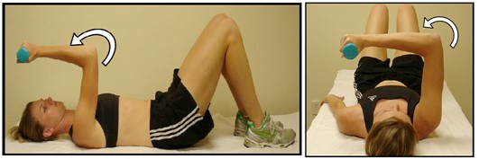
Our right triceps become very challenged if we can not sufficiently engage our left internal obliques and transverse abdominals during active late left lower extremity ‘push-off’ and concomitant active late right upper extremity ‘push-back’ (shoulder extension). Our bodies lose their ability to become centered over the left when our left abdominals, left hamstrings and right triceps disengage as a functional group. A few weeks ago Sy Abe-Hiraishi, a PRI faculty member, asked me about a non-manual technique called the ‘Supine Weighted Tricep Curl’ and the reasoning behind the two methods of instruction. I absolutely loved the question, the dialogue and the timing, since I will be presenting information on group function afferentation at this year’s Spring Symposium. Please read her summary and the response that is presented from a question that was initially asked by a course attendee that attended one of her courses in Japan. So awesome!   

Gratefully, Ron

アメリカの皆さんこんにちは!

Hello everyone!

日本講習担当、PRI講師の阿部さゆりです。昨年の12月に日本で初めてポスチュラル講習の開催をすることができたのですが、その際に参加者さんから出た質問とその答えを今日はここで共有したいと思っています。ロンが日本語と英語の両方でアップして!というので、両方でお届けしますですよ。

This is me, Sy writing this blog post – those of you who do not know me, I am one of the two PRI faculty members that are designated to teach courses in Japan. We hosted our very first Postural Respiration courses in Japan in December, and this interesting question came up from a participant. Today I am hoping to share the question and answer with y’all…(in case you cannot tell, I currently live in Texas). Ron specifically requested that I post this both in Japanese and English, so here it is – hope y’all enjoy!

ポスチュラル講習では左のハムストリングを使って骨盤を起こすように、右のトライセップスを使って右肩甲骨を起こすことがいかに重要か、という話をしますよね。その流れで講習中スーパイン・ウェイテッド・トライセップス・カールズの解説と実技ラボをおこなったのですが、このエクササイズにはやり方が二通り(ポジションA vs B)あり、「このエクササイズのポジションの違いは何?どういう状況の場合、どちらを選ぶのが正解なの?」という質問が日本人参加者さんから出たのです。

In Postural, we learn to use the Right Triceps to “get the scapular up” just like we learn use our Left Hamstring to “get the pelvis up” in the Myokin course.  One of the exercises we demonstrated and practiced in our Postural courses in Japan was “Supine Weighted Triceps Curls,” and that’s when this question was brought up – “I see, in the manual, that this exercise can be performed in two different positions – the original (Position A) and the alternate (Position B). What is the difference and what are some of the reasons we should pick one over the other?”

                           Position A                                                                 Position B

What a great question! This got Ron super-excited because no one in the U.S. ever asked this question to him, and Ron and I had a great conversation about it over lunch the other day. So let me first share the short version of the answer – “You should always try Position A first. If it does not work, try Position B as this should be the “mechanically advantaged” version of the same exercise.”

最初に答えを書いてしまうと、どんな患者相手にもまず試すはポジションAです。このポジションでは、歩行時に左足で身体を前に押し出して、右手を前・上方に振り切った状態(=右立脚中期)からエクササイズが始まるんだ、とイメージするとわかりやすいかも知れません。ここからPropel (前進)するために次にすべきは右腕を振り下ろし、後方に振り切ることですよね。同時に左腕も前に振り上げれば、体幹の右回旋・骨盤の左回旋と一緒に右足のpush-offが起こり、左前方への体重移行が可能になります。

平たく言えば、トライセップス・カールズはこの右腕の「振り下ろし始め」を練習するエクササイズなのです。エクササイズ開始時のポジションでは右肘と右肩は共に屈曲位にあり、上腕三頭筋をisolate(隔離)するのに最適と言えます。肘と肩、両関節で伸長位に置かれたこの筋肉を、肘伸展を通じて収縮させることで「腕の振り始め」に真っ先に上腕三頭筋にスイッチを入れる感覚を体得できた人は、そのあとの「右腕の後方振り切り」はモーメンタムと広背筋らの協力を得て比較的楽に、自然とおこなえるんだということに気が付くかもしれません。

And here’s the longer-version of the answer. Everything we do can be applied into various phases of the gait – so please picture yourself being in the right mid-stance. Your L AIC pushed you over to the right, you are feeling the right heel and the right mid-foot, and you just finished swinging your right arm all the way forward. Now look at Position A – both right shoulder and elbow are flexed – doesn’t it look awfully similar? If you are thinking this position is mimicking the I-just-finished-swinging-my-right-arm-forward phase, you are absolutely right!

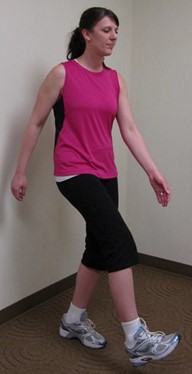
Now think what needs to happen next. You will need to begin swinging your right arm towards the back as you start to prepare yourself for the push-off phase, and eventually to shift your body weight to the left. The initiation of the right arm swing is essential because, without this, your right arm won’t be pulled all the way back, your right trunk won’t be pulled back, and the right trunk rotation won’t occur. Combined with L AF IR, the back-swing of the right arm is the key for the upper and lower body integration as shown in the picture below.

 (89) Left Stance Interrupted Swing

**改めて言及しますが、右の腕を後ろに引く、ということはロンがどの講習でも何度も繰り返し強調する、超超超超超重要事項です。右の腕を後方に振り切るということは右の体幹を後ろに引く、つまりは体幹が右に回旋するということでもあります(例: 下の写真参照)。体幹の右回旋は骨盤の左回旋(L AF IR)と対になるべきPRI介入には欠かせない要素です。ロン曰く、「左スタンス時(下写真)に荷重されていなければならない肢がふたつある。なんだかわかるかい?左足と、右腕だよ!」。歩行時に腕を荷重だなんて、その表現の仕方がまたロンらしいですけれど、つまるところ彼は空間把握や自我確立を導く神経的道具として、この状況では左足右腕が真価を発揮しているべきだと言いたいのです。

And hence the beauty of this exercise. Triceps curls in this position (Position A) is a great way for us to practice the initiation of the right arm swing. In this position, the triceps are elongated through the combination of shoulder flexion and elbow flexion and can be effectively isolated isolate.

しかし、このポジションで陥りやすいワナは「非常に矢状面に特化したエクササイズであり、上腕の屈曲には腰椎の伸展も伴いやすい」という点です。PRIの基本は適切なポジションで適切な筋肉を使う練習をするところですよね、ですから、患者さんがもしこのエクササイズで呼気(state of exhalation)のポジション、つまり胸郭の屈曲を保てなければ次はAlternate Position(代わりとなるポジション)であるポジションBを試すべきです。

However, here’s a pitfall of “Position A.” Because performing triceps curls in this position promotes sagittal movements, some patients just cannot help but extend their back. Those individuals weren’t quite ready to truly isolate the triceps and may need to take a step back – and try the alternate position, Position B.

ポジションBでは同じ歩行は歩行でも腕を縦ではなく横に振りながら、水平面で胴体を回旋させながら歩行してるイメージです。右の腕をぶん、と横にスイングし前に持ってきたようなこのポジションでは腰椎の伸展は格段に起こりにくいばかりでなく、体幹の左回旋が促進され、左腹壁の活性化と左後方縦郭の拡張、そして右肩甲骨の安定が起こりやすいのが特徴です。つまり、こちら(ポジションB)のほうが上腕三頭筋を単独で活性化させる(ポジションA)前に、腹壁・胸郭・肩甲骨と腕との統合をまずマスターしなければいけない患者に適したエクササイズなのです。

So now look at Position B. You also see the same gait phase in Position B, but the arm is swung to the side (on the transverse plane) instead of up to the front (on the sagittal plane). This arm position promotes the trunk rotation to the left, the better left abdominal engagement and the right scapular stabilization on the thorax, which further secures the right triceps. In this position, the patient will be given the mechanical advantage to remain in the state of exhalation (and thus not to extend the back) while activating the right triceps.

要約すると、「右上腕三頭筋の活性化にトライセップスカールを用いる場合、最初はポジションAで、もし腰椎の伸展がどうしても起こってしまう場合はポジションBでこのエクササイズをおこなう」ということです。こんな話をロンとしていたら、「こんな質問、アメリカでは受けたことないよ!」と非常にうれしがっていました。日本の方の思考力の助けを得ながら、これからも日本での講習でPRIコンセプトをこれでもかというほど切り刻んでいきたいと思います。アメリカの皆さんもこの回答を楽しんでいただければ幸いです。

Let me summarize it – “When using triceps curls to facilitate the activation of the right triceps, anyone should try Position A first. If the patient extends the back in this position, try the alternate position, Position B for the better abdominal integration.” I hope that this answer helps y’all, PRI families in both Japan and the U.S.!

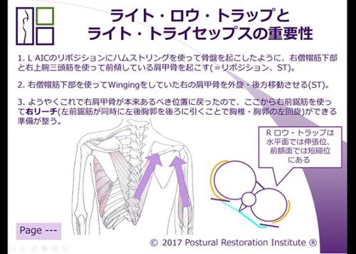
追記ですが、ロンに日本のPRI講習で使っているスライドを見せたら「なにこれ!すごいねすごいね、これも一緒にアップしてよね!」と興奮して大騒ぎされてしまったので、私たちが日本ポスチュラル講習で使ったスライドの写真もおまけに付けておきます。アメリカの皆さんにも、PRIを日本語に訳すのがどれだけ大変だったか、これを見れば少しだけわかっていただけますでしょうか?

P.S. – I showed this (power point slides that we used in Japan) to Ron and he told me I HAVE to post this photo in this blog. I created some visuals and added them to the slide explaining the importance of the right low trap and right triceps…well, I am sure y’all can read the rest ;)  We will continue to strive to teach high-quality, full-of-fun PRI courses in Japan!

Circle Explanation: That’s the cross-sectional view of the R BC-patterned chest….L lungs are more inflated than R, and the sternum tipped to the R. Orange semi-circles in this diagram represent SAs, and the light-blue lines are the lower traps. I use this diagram to explain how "L SA is rounded yet elongated (= challenged)" and "R SA is shortened yet flattened (= also challenged)" – sort of like obturator internus. Also, this diagram may help visualize how low traps are positioned in the transverse plane. For example, R low trap is shortened in the frontal plane (as shown in the image on the left…the back view), yet it is actually elongated in the transverse plane if you are to take a look at the image on the right (light-blue line)…the opposite is true for the L low trap… and therefore, again, both lower traps are challenged in its own way.

阿部さゆり

Sayuri "Sy" Abe-Hiraishi, MS, ATC/L, CSCS, PRT, NASM-PES, CES

Recent course question: Is the Asymmetrical Tonic Neck Reflex (ATNR) ever fully integrated, or is it constant presence throughout our lives?

The ATNR typically is inhibited by the age of 3.5 years. However when a lack of alternating head, trunk and appendage movement occurs, because of visual, auditory or tactile sensory orientation that reinforces homo-lateral movement, the retention of this primitive reflex can have an impact on behavioral formation of the body on rotation indefinitely.

Some of the retained symptoms include postural imbalance when the head is turned, difficulty in cross pattern movement of the trunk where one arm needs to move toward the controlled leg, hand-eye coordination difficulty over focus attention of the visual system, visual perception difficulties, cognitive learning challenges, excessive wrist and ankle tone visually on the right side, and bilateral functional integration difficulties in general.

The ATNR, like other reflexes that are considered to be associated with vision can be triggered or stimulated reflexively to some degree anytime in life when bilateral or alternating function is challenged by over referencing homo-lateral anti gravitational or positional strategies.

I am reviewing the Cervical revolution text and have a question. Page 46 in the grey box first point: Lateral Pterygoid pulls ipsilateral base of sphenoid down and forward while the ipsilateral (what the text states) OR contralateral (what seems correct) greater wing simultaneously raises? 

In OA osteo kinematics section the O rotates contralaterally to lateral flexion, Atlas goes opposite and Axis follows O (p31.) But in Arthrokinematics section Lateral flexion of O on A with conjunct contralateral O rotation, alar ligaments causes ipsilateral axial rotation and contralateral atlas rotation (atlas follows O.) Can you explain this? I cant seem to make it right in my head. 

Page 46 is correctly written and holding a model of a sphenoid or looking at a 3-D image of one would possibly help understand the relationship of the lower base of the sphenoid to the greater wing.   I can appreciate the difficulty you may have in their relationship.

The first sentence on page 31 relates only to Occipital (O) movement on the Atlas (A) in lateral flexion if the axis of the spine in general is in a neutral position. Often this is not the case. In a neutral position the last sentence on page 31 is also correct.  However, keep  in mind the mechanics of C1, or the atlas, on C2, or the axis, or C2 movement on C1 could  be challenged in lateral flexion if the lateral flexion of O on A or A on O occurred on a cervical spine that is positionally  laterally flexed to the right or to the left.

In the Arthrokinematic section of the Cervical Revolution manual, page 37 compliments page 31.  The top of page 38 is also correctly written with respect to the alar ligament and soft tissue in general.  Our control and stability of the O on A and the A on O depends on this counter balance provided by this tissue to offset osteokinematic momentum and obligatory osseous directive force.  Therefore, without this contralateral arthrokinematic tension the osteokinematic directional guidance from the bone surfaces of the O and A would promote over lateral flexion, rotation, sagittal movement at some place or point in the transverse or frontal planes and possible joint subluxation.

I hope this helps in understanding the role eccentric ligament and muscle tension plays during opposite obligatory osseous directed movement.  Thanks again for inquiry.   

Ron

For the Supine Active Sacro-Spheno Flexion, I have a few questions:
1. What is the benefit of the forward jaw?

2. Why not allow OA flexion? I thought the TMCC pattern involves SB flexion with OA extension and lower cervical flexion, wouldn’t the OA flexion promote the SB extension that is needed?

3. If we are using the Styloid to help ER the R temporal bone, where is the tongue (as it relates to the hyoid bone) during this exercise?

The benefit of moving the jaw forward is that the lateral pterygoids help move the cranium back and around the rolled up towel under the mid neck.  Cranial retraction allows more freedom at the articulating paired bones of the cranium and promotes OA extension, which is a precursor for cranial flexion (and is the expansion and “repositioning” segment of the technique).  The right TMCC pattern includes left SB flexion and OA extension when compared to the position of the right cranium that is in a probable state of right cranial extension and OA flexion. So, we are setting up for cranial flexion through the OA by acquiring “normal” OA extension or neutral position by bringing the mandible forward so that both sides of the cranium and cranial base are capable of being repositioned.  OA flexion would promote SB extension and if you would only perform this on the left, you would also promote more OA extension on the right when considering the influence the left OA would have on the right OA, during this accompanying cranial rotation to the right.  So, we want to promote more right OA extension in most humans to reverse or establish head or sphenoid rotation to the right and accompanying thorax rotation (T4) to the right .  I am using the styloid muscle to assist with ER and anterior rotation of the right temporal bone as OA extension is preserved across both occiputs and as cranial flexion is enhanced through right temporal ER.  The OA extension on the left should not interfere with cranial function since both sides of the cranium are now in flexion.  One would need more OA flexion on the left for left sphenoid or cranial extension, if more active cranial and head rotation to the right is desired.  But, in this technique we are establishing neutrality at the base of the head and then promoting biased cranial flexion on the right through temporal movement.  The tongue should be relaxed and inactive as it rests in the freeway space of the anterior oral cavity.  I am depending on the styloid hyoid and the strylopharyngeal muscle to pull the temporal styloid process and reposition the temporal bone on the right, not necessarily the styloglossus muscle, because the aforementioned muscle is now adhered to secured pharynx and hyoid because of the neutral rest position that the head and neck are in.  Too many of us use the styloglossus muscle to “move” or stabilize the temporals and this technique more than likely reduces the demands on the tongue and placing more demands on temporal pharynx and temporal hyoid movement versus pharynx temporal, hyoid temporal and glossus temporal  movement. 

Ron Hruska

Is it correct that the Left diaphragm is more responsible to pull the air to Right chest and Right diaphragm is more responsible to pull the air to Left chest? I was looking for the mechanism of this from PRI textbook and other resources, but could not find the one that makes this clear for me. I would really appreciate if you could explain the mechanism and/or give me some resource to explain that.

There is really no ‘one’ excellent resource to go to that will talk about hemi-diaphragm activity influence on the chest wall uniformly or non-uniformly.  There is no discussion how tidal volume or residual  volume of the lung is influenced by thoracic rotation or hyperinflation more so on one side of the chest wall than the other, even when you review the Scoliosis literature.  However, there is a great deal of information on how ribs are influenced by spinal coupling and directional rotation.  The costal aspect of the diaphragm is responsible for rib external rotation upon diaphragm contraction, while the crural fiber is primarily responsible for dome descension and thoracic lumbar spinal extension.  We have two sets of these muscles and when one rotates to the left with the rib cage or thorax, the ribs on the left are externally rotated with respect to the ribs on the right and the extension of the back at the thoracic lumbar vertebral wall is more extended on the left when compared to the right (Left AIC pattern).  Therefore, the costal and crural fiber on the left is at concentric end range and any respiration that occurs in this pattern will primarily occur with the hemi-diaphragm on the left because of the lengthened state of the crural and costal fiber on the right compared to the left.  Reciprocal breathing in this pattern will more than likely compliment reciprocal thoracic wall expansion on the right, because the costal fiber can pull the ribs up on this side, and the recoil upon exhalation is preserved because of the position the thoracic mediastinums.   Most researchers in pulmonary medicine or in functional performance do not measure tidal volume with individuals in this extreme state of torsion or rotation, either because they do not understand how posture can actually be challenged by these concepts or because it is a very untraditional method that would have a very difficult time being accepted by research reviewers and readers, since they probably would not appreciate the significance of thoracic mechanical expansion or lack of, when considering pulmonary function.  PRI is the only resource you will probably find that postulates these concepts that are scientifically sound because of what already exists in evidenced based research to date on rib mechanics, thoracic function and pulmonary studies.  Patterns of respiration such as the Left AIC or Right BC have not been ever considered to my knowledge.   

Asymmetrical ventilation and perfusion between the right and left lungs occurs in more than half of the children with severe congenital and infantile thoracic scoliosis.  However, the severity of lung function asymmetry does not relate to Cobb angle measurements.  Asymmetry in lung function is influenced by deformity of the chest wall in multiple dimensions, and cannot be ascertained by chest radiographs alone (Redding G. Song K. et al Lung function asymmetry in children with congenital and infantile scoliosis. The Spine Journal, 2008, vol 8,(4) 639-644.).  The last sentence describes it all.  It is very difficult to measure lung or perfusion function unilaterally with an x-ray, or any other pulmonary study method only because of the inability to separate flow from one side of the thorax upon normal non-compensatory postural positions versus compensatory postural positions that become neurologically habitual patterns of the “way” we breathe. 

Ron Hruska

How does the left low trap and serratus “secure the spine” (as mentioned for management of superior T4 following treatment of it)?

The left lower trapezius muscle rotates the mid thoracic spine to the right as it assists with internal rotation of the left ribs that are attached to the thoracic spine, and movement of the same ribs posteriorly. An individual with a superior T4 syndrome is experiencing difficulty in moving the right first and second ribs into internal rotation and the first and second ribs on the left, concomitantly, into external rotation upon left trunk rotation. During right trunk rotation, all the ribs on the left rib cage need to simultaneously rotate into internal rotation and likewise all the ribs on the right rib cage need to rotate into external rotation. After performing a right subclavius manual technique to increase the length of the right subclavius muscle, spinal rotation to the left is needed to take advantage of implementing internal rotation of all the ribs on the right, since now, ribs one and two on the right can internally rotate and ribs one and two on the left can externally rotate.

By reaching forward with the right arm to activate the right low trap and right serratus anterior for left spinal rotation and scapular movement on the thorax, you re-align all the ribs by moving the right ribs into internal rotation. Reaching forward with the right arm a little more upon exhalation would help this process even more. Then, reverse and move the left arm forward, upon exhalation, to rotate the spine and rib cage to the right. Every time you inhale, with the left arm reaching forward, expand or move back the left posterior ribs a little more. By moving the left ribs back on inhalation with the left serratus anterior, as the spine is being held or stabilized in right trunk rotation, the diaphragm can assist with air flow into the left posterior chamber of the left lung promoting universal posterior thoracic expansion during right trunk rotation without losing internal rotation provided by the left serratus anterior of the left lower ribs.

From this point on, your right lower trap will now experience more stabilization from the left lower and mid ribs and the left lower trapezius during alternating arm reaching or trunk rotation. In essence, the left lower trap is now serving as an eccentric stabilizing force for left posterior thoracic expansion during left heel strike and left arm reach. Without the left lower trapezius and left serratus anterior functioning upon left arm reach, the spine would not need to rotate to the right with the left lower trapezius because it may already be oriented to the right because of lost left posterior thoracic expansion and inability to open the apical or posterior left lung and chest wall. The left first and second ribs staying in an internal rotated state, limits the ability to expand the upper posterior mediastinum or lung tissue. Therefore, following the right subclavius manual technique used for treatment of a superior T4 syndrome, alternating right lower trapezius and right tricep with left lower trapezius and left serratus anterior is highly recommended for spinal stabilization during alternating trunk rotation or gait.

-Ron Hruska

More Clinical Questions